Technology
Microscopes and Imaging
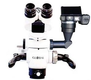 In our practice, an operating microscope with LED illumination is used during every endodontic procedure. With magnification up to 19.2x, we are able to provide the highest level of diagnostic and technical accuracy for your treatment. Our microscopes are also equipped with high-resolution digital imaging and video that we use in certain cases in order to provide you with important information concerning your tooth.
Digital Radiographs (X-rays)
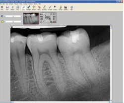 Digital radiographs have two key advantages. First, the radiation exposure is reduced up to 80% compared with dental film radiographs. Second, the digital image appears instantly on a wide-screen high-resolution monitor and is then integrated into your electronic medical record. You will be able to see image and your doctor will show you important features pertaining to your dental condition.
Nickel-Titanium Rotary Instruments
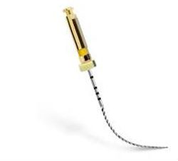
Rotary nickel-titanium files are the most efficient instruments for cleaning and shaping the root canal. These are used in conjunction with liquid antimicrobial agents to thoroughly disinfect the canal system.
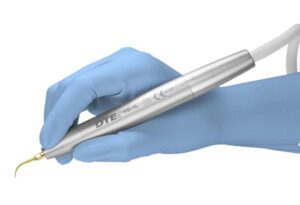
Sonic Activated Irrigation
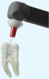 Applying sonic energy to our sterilizing solutions allows for more thorough cleaning of the root canal system. This device, which vibrates at 18,000 cycles per minute, helps remove debris and bacteria from areas that are difficult to reach with mechanical preparation alone.
Biocompatible Dental Materials

Bioceramics and Mineral Trioxide Aggregate (MTA) are dental materials that have many applications in endodontic procedures. Their biocompatibility and physical properties make these superior materials for endodontic microsurgical procedures (i.e., apicoectomy, surgical root repairs), as well as other endodontic treatment applications (i.e., endodontic treatment of the tooth with an immature root).
Ultrasonic Instruments
 Ultrasonic instruments, which vibrate up to 25,000 cycles per second, have multiple applications in endodontics. They are used to facilitate removal of restorative dental materials (e.g. posts, pins, and fillings) and for removal of canal obstructions (e.g. separated instruments and canal filling materials). Ultrasonic instrumentation also assists us in locating calcified canals, surgical root-end preparation, and disinfectant activation (to enhance cleaning the canal system).
Cone-Beam Computed Tomography
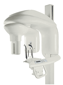
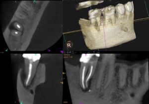 Our Cone-Beam Computed Tomography (CBCT) scanner uses state-of-the-art technology to take 3-dimensional images of teeth and surrounding structures. These 3-D images show anatomically accurate information that cannot be detected using conventional (2-D) x-rays. This information further aids our doctors in diagnosis and treatment planning for your dental condition. Our scanner takes limited field of view images of the area in question which reduces the radiation dose to the patient (equal to approximately 3 intra-oral x-rays).
See How CBCT Helps Endodontists Expose the Source of Tooth Pain
This video explains the benefits of using CBCT 3-D imaging in the diagnosis and treatment of dental disease. This video was produced by Dr. Kevin Kuo, endodontic resident at the Harvard School of Dental Medicine.
Root Canals Aren’t What They Used To Be
It’s not your grandfather’s root canal! Just like technology changes, so has root canal treatment.